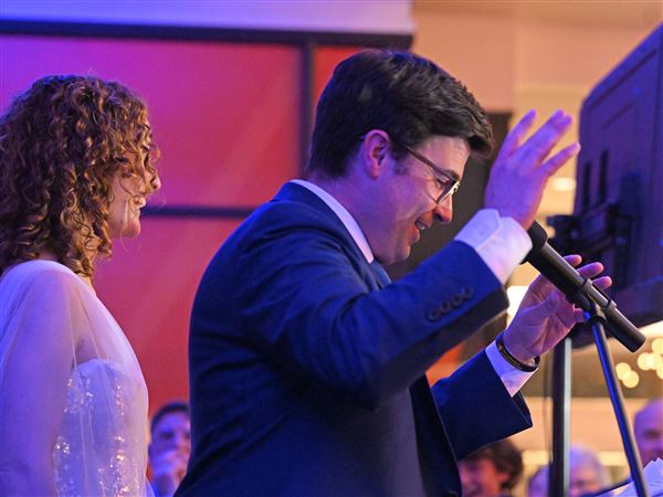
Even if it takes another 20 years to become a reality, Tim Hornik said, he would be willing to volunteer to get an eye transplant at the University of Pittsburgh.
Mr. Hornik, 35, was shot in the face by a sniper in Iraq in 2004. It destroyed vision in his left eye and damaged it severely in his right eye.
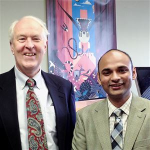
In the years since then, he has participated as a consultant for the U.S. Defense Department in reviewing research proposals on treating war injuries, including the futuristic effort being led by transplant surgeon Vijay Gorantla at Pitt.
Dr. Gorantla, who has helped pioneer Pitt’s hand and arm transplants, is the first to acknowledge that it will be years before surgeons can attempt whole eye transplants in human patients. But he says the approach has a key advantage over other attempts to repair traumatic injuries to the eye, whether they have come from a roadside bomb, an industrial accident or a car collision.
The eye is so complex that trying to repair its internal parts is an enormous challenge.
“The potential advantage of the eye transplant is you’re taking a whole eye with all its components, especially the retina,” Dr. Gorantla said. “You connect that to a residual optic nerve, if it’s present,” with the hope that the nerves from the donated eye will link up up successfully with the optic nerve going into the recipient’s brain.
Trained as a transplant specialist, Dr. Gorantla said he has “made an attempt to learn the intricacies of ophthalmology over the last five years, and I have phenomenal [research] fellows coming from across the world trying to learn about these things as well.”
At this point, the group’s work has involved rats and pigs. Colleague Kia Washington, a plastic surgeon, has transplanted whole eyes in rats and kept them viable for up to 200 days. While she has connected the eyes successfully, the group hasn’t yet done behavioral tests to see what kind of vision they provide to the rats.
No eye transplants have been done in the pigs yet, Dr. Gorantla said. First, the group is exploring the best surgical approaches for gaining access to the eye socket. The advantage of using pigs is that their eyes are only about 15 percent to 20 percent smaller than human eyes and the blood vessel connections are very similar, so “we can take bigger leaps in understanding what the challenges are in translating it to the bedside for humans.”
One of the biggest potential barriers with eye transplants is that the retina in a donated eye will die off within two to four hours of being separated from its blood supply. That will put a premium on the donor being near the recipient, Dr. Gorantla said, and, as with face transplants, it may work best if the donor is brought into the same operating room as the recipient.
In fact, for soldiers or others who have received devastating facial injuries, it might make sense to do a face and eye transplant at the same time, from the same donor, he said.
Another hurdle: The standard organ donor who has been declared brain dead may not work for eye transplants, because damage to the brain may harm the pathways from the eye to the brain’s visual centers. That could mean trying to find eyes from the much smaller pool of “cardiac death” donors, whose organs are taken immediately after the heart stops beating.
Even if those challenges can be overcome, “you have to be able to connect the retina through the optic nerve to the pathways to the brain, and there are 1.3 million nerves in the optic nerve.” said Randy Kardon, a traumatic eye injury specialist at the University of Iowa and the Iowa City VA Medical Center.
In addition, Dr. Kardon said, the patient’s immune reaction to a foreign eye could run the risk of interfering with those nerve transmissions.
Compared to other organs and tissues, though, the eye has some biological advantages that make it less likely to be rejected by the recipient, Dr. Gorantla said, and that might mean patients won’t need as strong a dose of anti-rejection drugs, and the drugs could be put directly into the eye.
Despite all these obstacles, Mr. Hornik, the injured veteran, is a pragmatic optimist about the procedure.
It wouldn’t bother him to take anti-rejection medication in return for regaining some of his sight, “and if it doesn’t work, they can just take the eye out.”
He spoke while wrapping up training with his new guide dog in New York City last week.
Now pursuing a doctorate in occupational therapy at the University of Kansas, Mr. Hornik can still vividly remember the day he was shot.
He was in a Bradley Fighting Vehicle and had his head exposed because the sand in the Iraq desert had fogged up the optical devices he normally used. The sniper’s bullet entered his left temple, and “it felt just like a sledgehammer hitting my head. I do remember my glasses beginning to separate from my head in milliseconds.”
Crumpled at the bottom of his vehicle, “I immediately began to assess the situation and was able to put my finger in the hole on the left side of my head,” but was confused because medics were bandaging his right eye. He later found out that’s where the bullet had exited. The bullet severed the optic nerve of his left eye but didn’t completely destroy his right eye.
After he recovered, he regained much of the vision in his right eye for a while, but he then went through a series of retinal detachments and cornea transplants, and now vision in that eye is down to 20/800. “For daily functioning,” he said, “I’m now down to shadows and light.”
After leaving the Army in 2011, Mr. Hornik got a master’s degree in social work at the University of Kansas, and now is working on a doctorate. His dissertation will focus on how people’s sense of identity changes when they become disabled in mid-life. To get through his schoolwork, he uses an Apple program that translates text into speech, and for the rare times he has to read a handwritten note, he’ll use a magnifier.
While the Pitt eye transplant program is decades away from fruition, Iowa’s Dr. Kardon is focusing on lower-cost techniques that might help veterans more quickly.
One technique his team is working on is a way of using light to quickly test the status of a damaged eye. If an injured eye is filled with blood and its pupil doesn’t contract normally in response to light, but the opposite eye’s pupil does contract normally when a light is shone into it, then a doctor would know the injured eye’s retina and optic nerve might be damaged, he said.
Dr. Kardon is hoping that approach can be incorporated into a low-cost, portable device that could be used at the scene of an injury. By comparing the pupil’s response to different wavelengths of light, the device may also be able to differentiate between retinal or optic nerve damage, he said.
Another approach he is excited about is the possibility of using telemedicine so that surgeons in the field could repair eyes with the help of an eye specialist sitting thousands of miles away, possibly with the aid of robotic surgery devices.
For longer-term therapies like eye transplants, Dr. Gorantla said, it’s important to be cautious and steady.
“We don’t want to create false hopes,” he said. “We want to say this is a very staged, conservative, thoughtful approach that goes from rats to larger animals and then will be translated at a point where we can be confident we can do this in humans.”
On the other hand, he doesn’t doubt the life-changing importance of what his group is attempting.
“For all of us, loss of vision is like losing ourselves, and for some of us, it is like death. Congenital blindness is different because you grow up with it and you build accommodating strategies. But for an adult to lose vision, it’s extremely challenging. If you say to most people I can give you the possibility to regain 30 percent of your vision just to regain some independence, they will say yes.”
First Published: July 6, 2015, 4:00 a.m.


